Lumbar Hernia

Lumbar disc herniation is a common spinal problem that occurs when the discs between the vertebrae press on the nerves and seriously affects the quality of life. It can cause severe back and leg pain (sciatica). Prof. Dr. Mehmet Tezer is a spine surgeon who specializes in all processes from herniated disc diagnosis to the most up-to-date surgical treatments. It is possible to get rid of these pains with early and correct treatment.
What is Lumbar Disc Herniation?
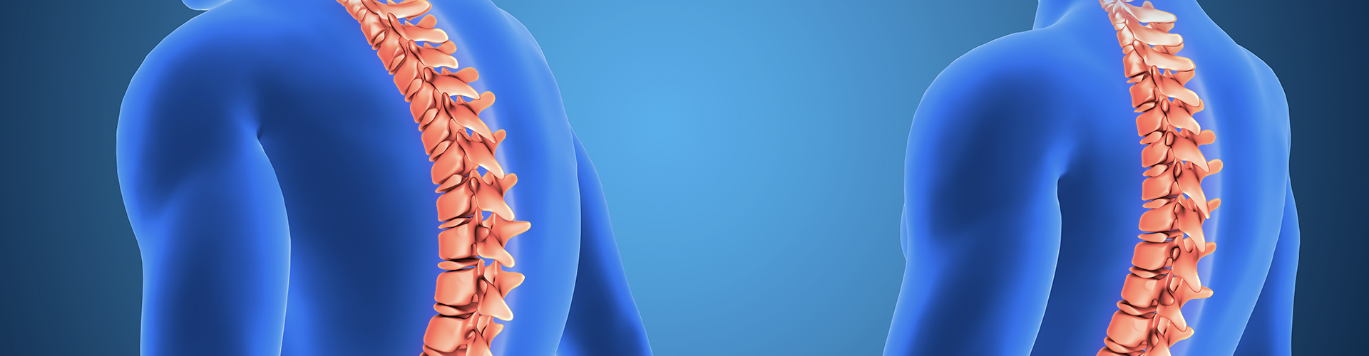
Our spine is made up of bones called “vertebrae” and “disc” structures that act as a cushion between these bones. These discs consist of a hard outer layer (annulus fibrosus) and an inner jelly-like center (nucleus pulposus).
A herniated disc is when the outer layer of the disc ruptures and the inner jelly-like part protrudes as a result of heavy lifting, forceful movement, trauma or age-related wear and tear (degeneration). This protruding disc fragment presses on the nerve roots passing through the spinal canal or going to the legs. This pressure is the main cause of pain and other neurological complaints.
What are the Symptoms of Lumbar Hernia?
The symptoms of a herniated disc depend on how much pressure the herniated disc is putting on which nerve. Although not all low back pain is herniated, the following symptoms indicate a typical herniated disc:
-
Pain in the Leg (Sciatica): It is a sharp, electric shock-like pain, usually unilateral, starting in the buttock and radiating to the back or side of the leg, sometimes to the foot. This pain may increase with coughing, sneezing or straining.
-
Low Back Pain Blunt or sharp pain that increases with movement and decreases with rest.
-
Numbness and Tingling: Pins and needles, numbness in the leg, foot or fingers.
-
Muscle Weakness (Loss of Strength): Weakness in the foot (e.g. inability to stand on tiptoe or heel) or leg, depending on the level of the nerve compressed by the hernia.


Diagnosis and Treatment of Lumbar Hernia
The diagnostic process begins with a detailed physical and neurological examination by Prof. Dr. Mehmet Tezer. Your physician evaluates the character of your pain, your reflexes and muscle strength. Magnetic Resonance Imaging (MRI) is the gold standard method for definitive diagnosis and to clarify the level, size and relationship of the hernia to the nerve.
Treatment is planned according to the patient's condition. The majority of patients can be treated with non-surgical (conservative) methods. These include short-term bed rest, painkillers and muscle relaxants, physical therapy and rehabilitation.
When is Lumbar Hernia Surgery Necessary?
Surgical treatment is considered in cases where conservative methods have failed or in cases of severe neurological problems. The decision for surgery is made in the following cases:
- Severe pain that does not go away despite non-surgical treatments (6-8 weeks on average).
- Significant and progressive muscle weakness (loss of strength) in the leg or foot.
- “Incontinence of urine or feces, known as ”Cauda Equina Syndrome" (this condition requires emergency surgery).
Today, herniated disc surgeries are performed with modern and minimally invasive techniques such as “microdiscectomy”. With this method, the herniated disc compressing the nerve is safely removed.
Frequently Asked Questions
No, no, no. Bel ağrılarının çok büyük bir kısmı (%80-90) "mekanik bel ağrısı" olarak adlandırılan, kasların zorlanması, spazm veya duruş bozukluklarından kaynaklanır. Bu ağrılar genellikle istirahatle ve basit ağrı kesicilerle birkaç gün içinde hafifler.
The main symptom suggestive of a herniated disc is pain radiating down the leg (from the buttock, back of the thigh or side of the leg to the knee or even the foot). In this case "sciatica" pain. If the pain in the leg is accompanied by numbness, tingling or muscle weakness, the suspicion of a hernia increases and a specialist should be consulted.
Yes, it can. A significant proportion of herniated discs, especially in the initial stages or small herniated discs, can heal without the need for surgery. The body's own defense mechanisms can shrink or "dry out" the herniated disc fragment over time.
The aim of non-surgical treatment is to support this process. Short periods of rest, anti-inflammatory medication, muscle relaxants and physical therapy and rehabilitation The programs aim to reduce pressure on the nerve and strengthen the muscles around the spine to speed up recovery.
The gold standard in the diagnosis of a herniated disc is to listen to the patient's complaints and take a detailed physical/neurological examination is to do this. Your doctor will check your reflexes, muscle strength and sensory loss.
However, to clearly see the location and size of the hernia, which nerve root it presses on and the degree of pressure Magnetic Resonance (MRI) imaging is essential. X-rays primarily show the bone structure; they do not show the disc, nerves and soft tissues. Therefore, MRI is the most reliable method in the diagnosis of hernia.
The decision to operate has less to do with the MRI image of the hernia and more to do with the patient's clinic, i.e. the symptoms they are experiencing. Surgical treatment becomes mandatory in three main cases:
-
Emergency (Cauda Equina Syndrome): Urinary or fecal incontinence, loss of sexual function or sudden and rapidly progressive paralysis of the legs. This is a rare but very serious condition that requires urgent surgical intervention.
-
Progressive Neurological Loss: Muscle weakness in the patient's leg or foot (e.g. inability to raise the ankle or tiptoe) that is marked and worsens over time.
-
Resistant Pain Severe leg pain (sciatica) that does not go away and disrupts the patient's quality of life despite trying non-surgical methods such as medication, physical therapy and injections for at least 6-8 weeks.
This is the biggest concern of patients. Today, "Microdiscectomy" surgeries (using a microscope or endoscope) are the gold standard in the treatment of herniated discs. With these techniques, the surgical field is magnified and the nerve tissue is clearly seen.
The risk of serious complications such as paralysis (paraplegia) in herniated disc surgery when performed by an experienced spine surgeon using modern techniques (microsurgery) is extremely low (less than one in a thousand). Although risks such as infection, bleeding or nerve damage are theoretically present as with any surgical procedure, these rates have been minimized with current technologies.
Microdiscectomy, is a minimally invasive method used in herniated disc surgery. It is performed through a small skin incision (about 2-3 cm) using a special operating microscope. The microscope magnifies the surgical field 25-40 times, allowing the surgeon to see the herniated disc and nerve root very clearly.
Advantages:
- Damage to healthy tissues (muscle, bone) is minimal.
- Bleeding is minimal.
- Postoperative pain is less.
- The hospital stay is short (usually 1 night).
- Recovery and return to normal life is much faster.
After microdiscectomy surgery, patients are usually up and walking 4-6 hours after surgery. They can be discharged the next day. Sciatica pain in the leg usually goes away immediately after surgery.
Patients are asked to be careful when sitting, standing and bending for the first few weeks. Desk work can usually be resumed within 1-2 weeks and heavier work after 6-8 weeks. In the postoperative period, exercise programs and physical therapy recommended by your doctor are critical for maintaining back health.
In herniated disc surgeries, the protruding disc fragment pressing on the nerve is removed, not the entire disc. There is a possibility that the remaining disc tissue may herniate again in the same place (recurrence). This rate is around %5-10 in operations performed by experienced surgeons.
The most important way to prevent hernia recurrence is lifestyle modification after surgery. Weight control, quitting smoking, paying attention to heavy lifting techniques and regular exercise, especially to strengthen the waist and abdominal muscles (core muscles), significantly reduce the risk of recurrence.